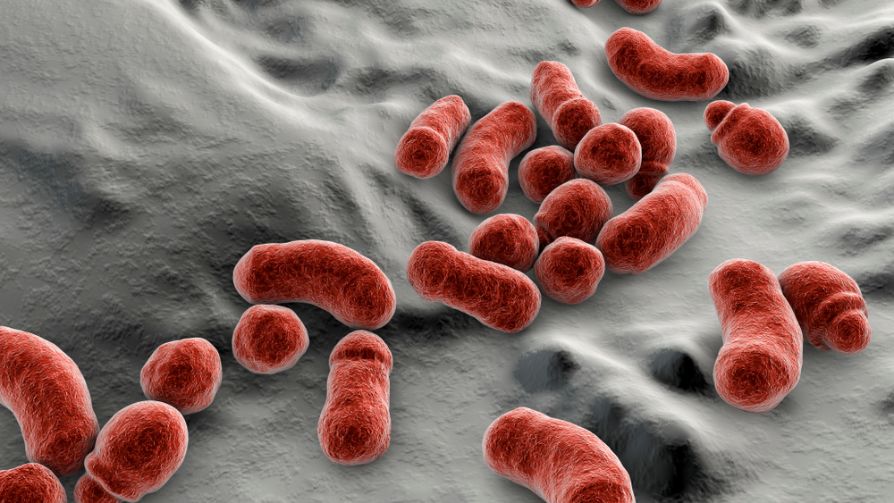
Malassezia fungi are naturally found on the skin surfaces of many animals, including humans. Various Malassezia species are also common in people. However, you can get a skin infection from a Malassezia fungus. Here's how to deal with it. Listed below are three important steps to cure eczema caused by Malassezia fungus.
Molecular methods for the reliable isolation of closely related Malassezia species
Several molecular methods have been developed and tested to differentiate closely related Malassezia species. PCR-RFLP analysis is one of these methods. DNA-targeted primers can be used to differentiate Malassezia species, which requires less expensive and technical equipment. It is also easier to perform than most molecular methods. However, the results of PCR-RFLP analysis cannot be relied upon for species identification.
PCR and restriction endonuclease analyses are two methods commonly used in clinical laboratories to identify Malassezia species. A total of 78 strains were analyzed to determine their variation in the number of ribosomal DNA repeat units. The products of the PCR reactions were digested using restriction endonucleases and the patterns generated were used to differentiate five of seven Malassezia species. In addition, M. sympodial yielded an amplified fragment of 700 bp, while the other six species had a greater sequence identity of 800-bp.
PCR-REA analysis has also been shown to be helpful in identifying different species. The ITS region of M. sympodialis can be differentiated from other Malassezia species using PCR-REA, while M. slooffiae and M. globosa show distinct restriction patterns. PCR-REA can differentiate closely related Malassezia species in as little as three cases.
Recent advances in research have led to a partial explanation of the role of Malassezia spp. in skin diseases. Genetic analyses are able to identify distinct genotypes within a single species. To ensure the reproducibility of molecular tests, reliable biochemical and physiological methods are required. These methods need to be accompanied by standardized molecular processes. When combined, these methods should yield reliable results for species identification.
PCR-RFLP is a rapid and accurate method for the identification of Malassezia yeast species. DNA extraction from skin tissue can be completed in less than 15 hours after the analysis. PCR-RFLP is widely used in dermatology laboratories and can be performed in under 15 hours. The results are consistent with the phenotypic and genotypic identification of the Malassezia species.
Molecular identification of a single specimen is important for identifying the species. The identification of closely related species is difficult using culture-based methods. Using the same ITS1/ITS2 sequence can differentiate closely related species. In addition, the same sequences have similar characteristics. Thus, the method can distinguish closely related Malassezia species. In this way, it is possible to identify an infectious genus without requiring a culture.
Incidence of Malassezia species on normal human skin
The incidence of Malassezia species in the microbiome of normal human skin is a complex phenomenon. Malassezia species are associated with certain diseases of the skin, and some are even pathogenic. In addition to contributing to skin disorders, these fungi may also trigger an immune response, causing various conditions such as type I allergy, eczema, and seborrheic dermatitis.
The colonization of the skin by Malassezia species is observed during the first few weeks of life. Neonates with high levels of M sympodialis are at a higher risk for infection, and the prevalence of the species was found in neonates from birth. These factors, as well as maternal contact, are likely to influence colonization. Several factors may be responsible for this, including sebum secretion and skin characteristics.
The incidence of Malassezia species varies in different parts of the body and may be influenced by other factors, including gender and age. The most common species were M. globosa, M. restricta, and M. globosa in adults aged 35 and older. However, these differences may be accounted for by differences in geographical specificity or diagnostic methods.
The incidence of Malassezia species in different parts of the body is more variable than in other regions, but the species are distributed in parallel to pilosebaceous gland activity in these areas. The density of these glands and the concentration of lipids vary between different parts of the body. The molecular analysis method has a higher detection rate, as well as conventional culture methods. This difference may be due to differences in sample collection methods and the different sebum compositions of individuals from different ethnic backgrounds.
The incidence of Malassezia species is associated with an imbalance in skin microbial homeostasis. The secreted metabolites from Malassezia may trigger the production of IL-17A by innate immune cells. Inhibition of these cytokines may contribute to the overgrowth of Malassezia on normal human skin. But the importance of the signaling cannot be overstated.
The incidence of Malassezia species in normal human skin is highly variable and dependent on underlying immunological conditions. It is important to note that the human immune system cannot completely eliminate Malassezia species from the skin. Therefore, it is important to understand that Malassezia colonizes the skin of normal human skin during the first few weeks after birth. It has been reported in as many as 30 to 100% of newborns.
One of the most recent studies on Malassezia has shown that in infected HIV patients, the incidence of Malassezia spp. is higher than that of healthy human skin. Interestingly, HIV-infected HIV patients are more likely to have severe SD, although this is rarely observed in healthy human skin. Furthermore, the prevalence of Malassezia spp. in HIV patients is significantly higher than in healthy Caucasian people.
Treatment of eczema caused by M. sympodialis
M. sympodialis is the dominant commensal fungus on human skin. It is associated with a variety of common skin disorders. This species produces exosome-like vesicles called “MalaEx,” which contain allergens and induce inflammatory cytokine responses. This fungus has diverse genetic features, which may contribute to its involvement in human skin diseases.
A mild soap should be used to wash the skin, and a moisturizing cream can help seal in moisture. Apply the cream at least twice daily and wash your clothing in lukewarm water rather than hot. You should also avoid wearing scratchy clothing and wearing loose, natural materials. Wearing new clothes should be washed first to ensure the eczema-causing organism does not spread to other parts of your body.
Some treatments for eczema caused by M sympodialis include antibiotics. However, steroid medications can have serious side effects, including bone loss. Other treatments include topical immunomodulators and ultraviolet light. Light therapy, which uses ultraviolet light to treat eczema, can also help. Light therapy, which uses special lamps to produce ultraviolet light, can help reduce the symptoms of eczema.
Among the treatment options for eczema caused by M sympodialis, re-establishing the epidermal barrier, and avoiding IgE sensitization are the most important targets. In addition to traditional topical steroids and immunosuppressive drugs, antifungal medications can also be beneficial. Topical calcineurin inhibitors are also commonly used.
In one case, a 67-year-old man with a history of multiple eczema lesions for more than two years was diagnosed with M. sympodialis after biopsies. The urine antigen test was negative for Histoplasma infection. Cultures of biopsies did not grow pathogens. The chest roentgenogram was normal. Morphologic examination of the skin lesions revealed that the patient had multiple infections of Malassezia sympodialis.
Systemic antifungals, such as metronidazole, are used to treat severe cases of M. sympodialis. These medications are also effective in cases of severe infection and treatment failure with topical agents. The antifungal medication will also help to prevent the yeast from multiplying in the skin, so topical treatments may not be enough.


Leave feedback about this