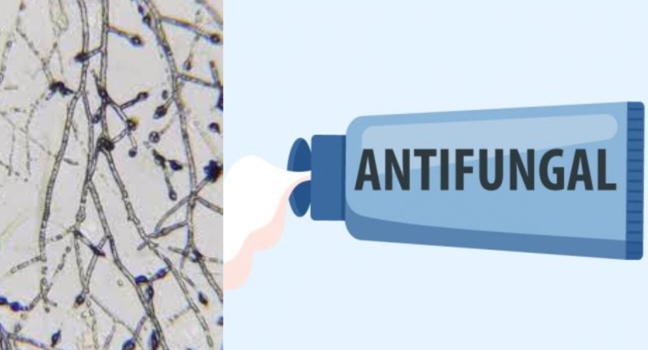
Dermatophyte fungi are obligate parasites on humans. Their infection causes significant morbidity and cost to society. They are grouped into three genera: Trichophyton, M. nanum, and T. ajelloi. Listed below are the key characteristics of each species and their treatment. In addition, this article will cover the immune response and phylogeny.
Phylogeny
Phylogeny of the dermatophytes shows that there are 3 genera: Trichophyton, Microsporum, and Epidermophyton. The two latter genera are paraphyletic, but Trichophyton diverged from Microsporum earlier than all other species. Its geophilic features may have helped it gain an ecological niche early on. The phylogeny of the dermatophytes reveals that Trichophyton species may have diverged from other fungi.
The phylogeny of dermatophytes is based on the Bootstrap values from 100 replicates. The red areas highlight dermatophytes and the black dots represent pathogenic fungi. The symbols next to fungal species indicate the host in which they grow. For instance, A. niger is mostly pathogenic to plants, but can infect humans if large amounts of spores are inhaled. By contrast, C. glabrata is only a problem for people with low immunity.
While a molecular approach to dermatophyte taxonomy has identified the major trends of evolution, it was unable to reliably classify the species. A pair of species was previously thought to be closely related, and it has been observed that the two strains are not related, although ribosomal markers can identify them. The multidirectional approach to dermatophyte taxonomy is necessary for accurate identification.
A recent study suggested that the AT-rich nature of the dermatophyte mitochondrial genomes may have influenced the third codon position. The two most common codons in the dermatophyte genomes favor A and T, while rarer codons have a greater G/C ratio. Thus, dermatophyte mtDNA is highly conserved across species.
Phylogeny of Dermatophytes has a strong relationship with the taxonomy of dermatophytes. This phylogenetic analysis provides a framework for understanding the biology of this fungus class. They are the most common fungal species found in humans, with a prevalence of between twenty and 25 percent. The etiological factors of superficial fungal infections are derived from dermatophytes. Moreover, they have remarkable adaptability to changes in environmental conditions. This makes them one of the oldest recognised pathogens.
The genomes of dermatophytes have the same order of conserved genes. It is also the case for other pathogenic filamentous fungi, including Aspergillus niger and Penicillium marneffei. The order of the conserved genes in the mtDNA of Candida albicans is largely identical among these fungi. It seems that the phylogenetic tree was able to capture this pattern.
Characteristics
Dermatophytes belong to the class of imperfect fungi, known as Deuteromycetes. They have a sexual stage and produce microconidia. Several other traits distinguish dermatophytes from other fungal species: they have undulant, branched hyphae with numerous septa along their lengths, which break off at the septa and produce amino acid monomers.
A dermatophyte fungus invades keratinized tissues and can cause infection. A common type is dermatophytosis. Symptoms of dermatophytosis include inflammation, alopecia, and hyperhidrosis. The fungus is found in soil and can be transmitted by contact or fomites. The organisms cause infections in a number of locations and cause a wide range of diseases.
Symptoms of dermatophyte infection are typically described as erythematous scaly plaque with a raised border. It may also be accompanied by a peripheral scale that points inward. Atypical infections are sometimes associated with kerion development and deep abscesses. In some cases, dermatophyte infections may be caused by non-dermatophyte fungi.
There are two main types of dermatophyte fungi: zoophilic and anthropophilic. Zoophilic species are closely associated with animals, and may cause a dermatophyte infection in humans. Both types may be transmitted by direct contact with an infected host or indirectly through infected shed skin and hair. A zoophilic dermatophyte can live up to 15 months before its virulence is reduced.
The other type of dermatophyte fungi is called homothallic. In this case, two types of reproductive structures (sex and growth) are required for reproduction. In humans, the most common species of dermatophyte fungus is Trichophyton rubrum, while in domesticated animals and pets, a different mix of dermatophyte fungi may be present. Treatment for ringworm depends on the type of dermatophyte fungus that is infected. Some common topical medications are fluconazole and griseofulvin.
The other type of dermatophyte fungi is trichophytonasis, which affects keratinized surfaces. Dermatophytes are commonly associated with ringworm or tinea. Dermatophytes are grouped by the site in which they are found. If they are associated with hair, they are referred to as tinea corporis, dermatophyte onychomycosis, or tinea unguium.
Treatment
The clinical cure of dermatophytosis depends on the type of infectious microorganism, the site of infection and the severity of the disease. Depending on the type and severity of the disease, treatment options may include topical and systemic antifungal agents. Prevention is essential to avoid the spread of the infection, as the condition can worsen if left untreated. A few preventive measures include avoiding close contact with infected persons and keeping personal items and clothing away from the infected area.
A wood's lamp examination involves shining a UV light on the infected area to determine if it is infected by dermatophytes. Unfortunately, this treatment option does not work for many dermatophyte infections. Nevertheless, it can help the doctor identify the type of dermatophyte and provide useful information regarding treatment options. Other tests include fungal culture and biopsy.
The most common fungus-causing a skin infection is the tinea pedis. In some cases, it can be difficult to differentiate between the two, so treatment options for tinea pedis are limited. Dermatophyte fungus can also be mistaken for a similar skin condition, such as acne. Fortunately, the symptoms of this condition are not always obvious, which may delay the diagnosis.
In most cases, the treatment of dermatophyte fungi involves topical antibiotics. Antifungal creams and lotions can be used to treat a dermatophyte infection. Topical agents are recommended to treat this fungal infection, but the fungi may be resistant to topical medications. A treatment for dermatophyte fungus should also address the underlying cause.
Increasing numbers of cases of cutaneous dermatophytosis are being reported worldwide, particularly in the tropics. The disease poses a considerable therapeutic challenge for dermatologists. Despite the numerous fungi, standard dermatology textbooks, which focus on dermatophytosis, have lost much of their relevance in the clinical setting. Dermatophytophytosis management in India requires an evidence-based practical approach.
Immune response
In immunocompetent hosts, dermatophyte fungi produce only superficial fungal infections, but they can infect immunocompromised hosts and cause invasive disease. The mechanisms that control the innate immune response to dermatophyte fungus infection are still unknown. To investigate these mechanisms, we examined damage caused by dermatophytes on epidermal tissues and primary keratinocytes. We identified signaling pathways and examined whether dermatophytes induce proinflammatory responses in both epidermal and keratinocyte cells. Invasiveness was induced by five dermatophyte species, with Microsporum gypseum causing the greatest tissue damage.
Several immunological pathways have been implicated in the dermatophyte response. The MAPK signaling pathway, which leads to the expression of the c-Fos transcription factor, has been identified in organotypic epidermal models. Furthermore, it has been shown that T. equinum induces expression of c-Fos, MKK1, and p38 kinase. In addition, it activates the expression of several transcription factors and signaling pathways.
The immune response to dermatophyte aphids is initiated by the production of activating cytokines by lymphocytes and other cells, including keratinocytes. In addition, keratinocyte activation is often correlated with damage induction. As damage occurs, tissue remodelling occurs. Activation of TIMP gene expression is reported for the first time.
Although the role of C-receptors in the cutaneous immune response has been questioned, the role of neutrophils has been confirmed. Neutrophils, which help the immune system recognize and eliminate foreign bodies, also help the body fight fungal infections. In vitro studies of C-receptors and neutrophils have revealed that these cells are involved in the defense against dermatophyte fungus.
Several studies have suggested that CLRs play a crucial role in the immune response to T. rubrum, and that their deficiency compromises the clearance of the fungal infection. The immune response to dermatophyte fungus, however, is not dependent on IL-17 response or adaptive immunity. This is an important factor for fungal control. But the key role of CLRs in fungal clearance is unclear.
A recent study investigated whether DC-HIL (a CLR for dermatophyte fungus) induces a specific cytokine response. This CLR had previously been associated with inhibitory signals that could inhibit T cell proliferation and the posterior reactivation response. However, it now plays a positive role in DC activation and cytokine secretion.


Leave feedback about this